Cell Stretcher
✓ Stretch cells or tissue mechanically
✓ Record & stimulate electrophysiological activity
✓ Optical or fluorescence imaging
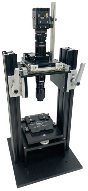
BMSEED’s MicroElectrode Array Stretching, Stimulating, und Recording Equipment, or MEASSuRE, is a complete solution for researchers to stretch cells/tissue mechanically, image them optically, and record/stimulate electrophysiological activity, separately or concurrently.
Each model of MEASSuRE integrates three distinct methods into one system: (1) a cell stretching device, (2) a data acquisition system for electrophysiology, and (3) a live cell imaging system.
The combination of these experimental paradigms is enabled by BMSEED’s proprietary stretchable microelectrode array (sMEA) technology, which contain elastically stretchable electrodes embedded in an elastomeric matrix that contact the cell or tissue culture, address this challenge by reproducing the mechanical and electrical environment of cells in vivo in a controlled environment in vitro.
Why Our Cell Stretcher?
✓ Reduced cost and complexity compared to other systems
✓ Real-time, in-situ, and label-free measurements
✓ Suitable for dissociated cell cultures, slices, and organoids
✓ Chronic and acute experiments
✓ Mimic biophysical in vivo microenvironment to more accurately predict in vivo behavior using in vitro data
✓ Electrically stimulate cells in culture
✓ Assess efficacy and toxicity of drugs
✓ Low-cost data acquisition and analysis
Our team of experts...
Founder
Phone: +1 (609) 532-9744
Email: oliver@bmseed.com
Application Fields of Cell Stretchers
(I) Physiological Cell Stretchers for Tissue Engineering
Pluripotent stem cells that differentiate into specialized cells have properties that more closely resemble adult tissue when the cells are mechanically and electrically stimulated during the differentiation using BMSEED’s cell stretcher, MEASSuRE.
MEASSuRE provides electrical and mechanical stimulation in conjunction with optical or fluorescent imaging.
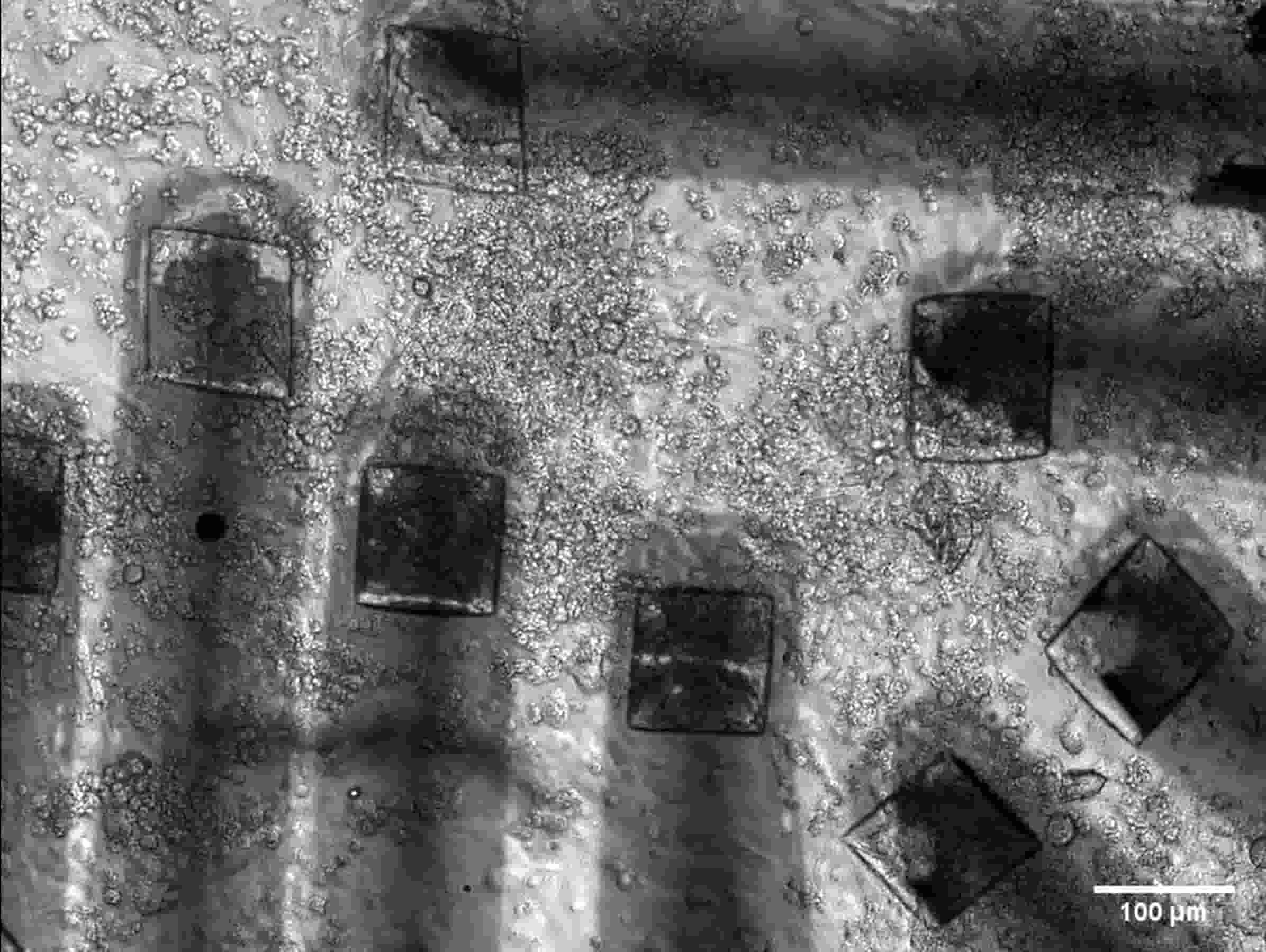
Image: hiPSC-CMs on BMSEED’s stretchable MEA, A. Patino Ph.D., Arizona State University
(II) Physiological Cell Stretchers for Drug Toxicity Testing
The validity of in vitro drug toxicity testing is increased when cells are derived from humans (not animals) to closely represent in vivo cells of the respective organ. This can be achieved by differentiating human induced pluripotent stem cells (hiPSCs) into specialized cells under mechanical and electrical stimulation.
MEASSuRE can be used as a physiological cell stretcher that provides electrical and mechanical stimulation in conjunction with optical or fluorescent imaging.
Image: hi-PSCs on MEA using BMSEED’s electrophysiology module & temperature controller, A. Patino Ph.D., Arizona State University
(III) Physiological Cell Stretchers for Mechanobiology
Biomechanics and modeling in mechanobiology play a crucial role in understanding the numerous mechanisms for transducing and sensing mechanical forces in neurons and other cell types, including those involved in cancer mechanobiology and cellular mechanobiology.
MEASSuRE can be used a physiological cell stretcher to understand the effect of mechanical forces on cell behavior.
Image: neurons on BMSEED’s stretchwell, A. Palmieri B.S., Georgia Institute of Technology
(IV) Pathological Cell Stretchers for Neurotrauma
BMSEED’s pathological cell stretcher, or MEASSuRE, can provide insights into neurotrauma by modeling the biomechanical forces that cause neuronal injury and comparing post-injury electrophysiology to their pre-injury levels. MEASSuRE can replicate the mechanical stresses involved in neurotraumas such as traumatic brain injury (TBI) and spinal cord injury (SCI), and assess how these stresses affect neuronal health and function. The effectiveness of neuroprotective treatments to minimize the damage after injury can therefore be readily assessed.
(V) Pathological Cell Stretchers for Concussion
BMSEED’s pathological cell stretcher, or MEASSuRE, allows researchers and physicians to develop improved concussion protocols that are based on the electrophysiology of the underlying injury rather than cognitive assessments. Researchers can develop more accurate and physiologically relevant concussion protocols by analyzing the changes in neuronal electrical activity following a concussion-like injury simulated by MEASSuRE. This can lead to improved strategies for diagnosis, treatment, and management of concussions as researchers learn about the injury mechanisms.
(VI) Pathological Cell Stretchers for Muscle Injury & Pain
BMSEED’s pathological cell stretcher, or MEASSuRE, allows researchers to investigate the mechanism of muscle injuries that are caused by excessive tension or compression, and the evaluation of drugs to speed up recovery. MEASSuRE can simulate these mechanical stresses and evaluate their effects on muscle cell health and function. Researchers can then test various treatments and drugs aimed at reducing muscle damage and accelerating recovery to develop therapies for muscle injury and chronic pain conditions.
(VII) Pathological Cell Stretchers for Stem Cell Repair Mechanism
Stem cells are involved in repair processes after injury in different parts of the body, e.g., in the brain after a traumatic brain injury. The mechanism of the activation of the mechanoreceptors is not understood. BMSEED’s pathological cell stretcher, or MEASSuRE, can be used to study how mechanoreceptors on stem cells are activated in response to mechanical stress, and how this contributes to the repair processes after injury. By simulating injury conditions and monitoring stem cell behavior, researchers can gain insights into the mechanisms that drive stem cell activation, differentiation, and repair to develop more effective regenerative therapies.
(VIII) Pathological Cell Stretchers for Alzheimer’s Disease
Neurodegenerative diseases such as Alzheimer’s disease (AD) have common pathological pathways with TBI, e.g., the build-up of amyloid-plaques. BMSEED’s pathological cell stretcher, or MEASSuRE, can contribute to AD research by replicating the pathological conditions similar to those observed in TBI, such as amyloid plaque formation. By using MEASSuRE to assess how these conditions affect neuronal function and health, researchers can evaluate the efficacy of new drug candidates in mitigating the effects of amyloid plaques and other AD-related changes in neuronal activity.
What Clients Say About our Cell Stretcher
“Tuneable parameters and very fast to use.”
- A. Pybus, Ph.D. Georgia Institute of Technology
“Easiness for acquiring electrical signal in a non-invasive manner.”
- A. Patino, Ph.D. Arizona State University
“The most valuable thing is that we can record from cells multiple times…injure and record on the same device.”
- M.K. Dwyer, Ph.D. Columbia University
Cell Stretching Systems
BMSEED’s MicroElectrode Array Stretching, Stimulating, und Recording Equipment, or MEASSuRE, is available in stand-alone modules or as an integrated cell stretcher, electrophysiology, and imaging system.
MEASSuRE Mini
Physiological Stretch Applications: The MEASSuRE Mini cell stretcher is ideal for research in tissue engineering, regenerative medicine, organ-on-a-chip systems, mechanobiology, and more.
Benefits of the MEASSuRE Mini cell stretcher include: 1) Physiological stretch of cells to reproduce the normal environment of cells inside the body to promote cell maturation, health and function; 2) Electrophysiology measurements with stretchable microelectrode array (sMEA) to assess cellular health, function, and maturity in-situ and real-time; 3) Imaging to visualize cells and cellular processes during stretch.
Key features of the MEASSuRE Mini cell stretcher include: 1) Mechanics Module: stretching at strain rates up to 1/s and strains up to 20%; 2) Imaging Module: optical or fluorescence imaging during stretching; 3) Electrophysiology Module: 2x60 channels for recording and stimulation
The cell stretcher offers precision with the smallest motion of 1.6 µm and the largest stroke (travel) of 6 mm.
Its compact design makes the cell stretcher most suitable for lab settings, including both incubator and benchtop workstations.
Contact us for pricing information.
MEASSuRE Premium
Pathological Stretch Applications: The MEASSuRE Premium cell stretcher is ideal for traumatic brain injury (TBI) and repeated concussions, spinal cord injury (SCI), neurodegenerative diseases, muscle injuries, etc.
Benefits of the MEASSuRE Premium cell stretcher include: 1) Pathological stretch of cells to reproduce the biomechanics of an injury such as traumatic brain injury (TBI) or spinal cord injury (SCI); 2) Electrophysiology measurements with stretchable microelectrode array (sMEA) to assess cellular health and function before and after the injury, with and without neuroprotective compounds; 3) Imaging to visualize cells and cellular processes during stretch.
Key features of the MEASSuRE Premium cell stretcher include: 1) Mechanics Module: stretching at strain rates up to 50/s and strains up to 50%; 2) Imaging Module: optical or fluorescence imaging during stretching; 3) Electrophysiology Module: 2x60 channels for recording and stimulation
The cell stretcher offers precision with the smallest motion of 6 µm and the largest stroke of 25 mm
Most suitable for lab settings like the benchtop
Contact us for pricing information.
MEASSuRE X
Pathological Stretch Applications: The MEASSuRE X cell stretcher is ideal for traumatic brain injury (TBI) and repeated concussions, spinal cord injury (SCI), neurodegenerative diseases, muscle injuries, etc.
Benefits of the MEASSuRE X include: 1) Very fast pathological stretch of cells to reproduce the biomechanics of an injury such as traumatic brain injury (TBI) or spinal cord injury (SCI); 2) Electrophysiological measurements with stretchable microelectrode array (sMEA) to assess cellular health and function before and after the injury, with and without neuroprotective compounds; 3) Imaging to visualize cells and cellular processes during stretch.
Key features of the MEASSuRE X cell stretcher include: 1) Mechanics Module: stretching at strain rates up to 90/s and strains up to 50%, 2) Imaging Module: optical or fluorescence imaging during stretching, 3) Electrophysiology Module: 2x60 channels for recording and stimulation
The cell stretcher offers precision with the smallest motion of 8 µm and largest stroke of 32 mm
Most suitable for lab settings like the benchtop
Contact us for pricing information.
Frequently Asked Questions About our Cell Stretchers
1. What is a cell stretcher?
A cell stretcher is a specialized laboratory device designed to apply controlled mechanical forces to live cells in culture. It mimics physiological stretching conditions that cells experience naturally in the human body, such as the cyclic stretching of lung alveolar cells during breathing, cardiomyocytes in a beating heart, or endothelial cells lining blood vessels under pulsatile blood flow. In addition, cell stretchers can mimic the pathological stretching conditions that cells experience during trauma, such as the impulse stretching of nervous tissue during traumatic brain injury (TBI) and repeated concussion or muscle tissue during excessive tension or compression. By simulating these biomechanical forces, cell stretchers allow scientists to study how cells respond to mechanical stress at a molecular, cellular, and tissue level.
Cell stretchers are critical tools in fields like:
Tissue engineering: Studying how mechanical cues influence cell growth and tissue formation.
Regenerative medicine: Investigating how physical forces impact cell regeneration and healing processes.
Organ-on-a-chip technology: Simulating organ-like conditions with mechanical stimulation to better mimic in vivo behavior.
Mechanobiology: Understanding how mechanical signals are converted into biochemical responses.
Drug testing and development: Observing how mechanical strain affects cellular drug responses.
By offering precise control over the magnitude, frequency, and duration of applied forces, cell stretchers provide invaluable insights into how mechanical stress regulates cell behavior, signaling pathways, gene expression, and overall cellular health.
2. What are the benefits that a cell stretcher provides over its alternatives (static cell culture, hydrogel-base models, etc.)?
Cell stretchers stand out as an essential tool in cellular and mechanobiology research due to their precision, versatility, and ability to replicate physiological and pathological conditions. While alternative methods like static cell culture, hydrogel-based models, or shear stress systems offer some insights into cellular responses, cell stretchers excel in providing controlled, repeatable, and physiologically relevant mechanical stimulation to cells with the use of stretchable microelectrode arrays (sMEAs). Below, we’ll explore the key benefits of a cell stretcher over its alternatives.
1. Physiological Relevance
Dynamic Mechanical Stimulation: Unlike static cultures or hydrogel models, our cell stretcher can replicate cyclic stretching, a critical mechanical cue experienced by cells in tissues like cardiac muscle, lungs, or blood vessels.
Accurate Simulation of In Vivo Conditions: Cell stretchers mimic strain rates, amplitudes, and durations observed in real biological systems, offering an accurate model for studying mechanical cues.
Targeted Stretching Patterns: Our cell stretcher allows for uniaxial, biaxial, or radial stretching with one single tool, whereas many alternatives lack this flexibility. Readily change the desired strain profile simply by switching out MEASSuRE’s indenter.
Example: Studying how cardiac myocytes align and function under cyclic stretching replicates the natural environment of heart tissue during a heartbeat.
2. Precise Control Over Experimental Parameters
Strain and Strain Rate Control: BMSEED’s cell stretchers allow researchers to fine-tune parameters like strain percentage (up to 50%) and strain rate (up to 90/sec).
Customizable Protocols: With software integration, BMSEED can help you design static, cyclic, or impulse stretching profiles for specific experimental requirements.
Repeatability: The mechanical stimuli programmed in MEASSuRE can be reproduced consistently, ensuring reliable results across multiple experiments.
Contrast: Alternatives like hydrogel matrices or shaking platforms lack the precision and repeatability of a dedicated cell stretcher.
3. Integration with Imaging and Electrophysiology Modules
Real-Time Imaging: BMSEED’s cell stretcher supports optical and/or fluorescence imaging during stretching experiments, allowing live visualization of cell morphology and intracellular processes.
Electrophysiology Integration: BMSEED’s stretchable microelectrode arrays (sMEAs) enable real-time recording of electrical activity in cells under mechanical stress. Researchers can readily compare pre- to post-stretch electrophysiology recordings.
Contrast: Static culture systems lack dynamic monitoring capabilities, while other mechanical stimulation devices may not integrate seamlessly with imaging or electrophysiology equipment.
-
A cell stretcher operates by applying mechanical deformation (stretching) to a flexible, cell-seeded substrate, such as BMSEED’s stretchable microelectrode arrays (sMEAs)made from PDMS (polydimethylsiloxane). The mechanics module is the heart of a cell stretcher, controlling the strain (how much the substrate stretches) and strain rate (how quickly the stretching occurs).
Stretching can be applied in different patterns:
Uniaxial Stretching: Stretching in one direction.
Biaxial Stretching: Stretching in two perpendicular directions simultaneously.
Radial Stretching: Stretching radially in all directions.
Stretching can be applied in different
Cyclic Stretching: Repeated stretching and relaxation cycles that replicate physiological mechanical cues.
Impulse Stretching: Single stretch pulses, often at high strain and strain rates to replicate injury.
Static Stretching: A static stretch at any strain for musculoskeletal applications, for example.
With BMSEED’s MEASSuRE, strain levels can reach up to 50%, and strain rates can go up to 90/second, replicating real-world cellular mechanical environments to traumatic cellular events.
The Cell Stretching Process in Action:
Cell Seeding: Cells are seeded onto the sMEA membrane and allowed to adhere and grow.
Experimental Setup: The sMEA is secured in the cell stretcher, MEASSuRE, with parameters pre-programmed in the software.
Mechanical Stretching: The device applies controlled stretching to the cell-seeded membrane to reach the desired strain at the desired rate.
Real-Time Monitoring: Imaging and electrophysiology systems capture cellular responses in real time to compare to pre- and post-levels.
Data Analysis: Data are analyzed to draw conclusions about cellular behavior under mechanical stress using MATLAB or NeuroExplorer, for example.
-
The cell stretcher can be used with a variety of cell types, including but not limited to:
primary hippocampal neurons
organotypic hippocampal slice cultures
organotypic spinal cord slice cultures
neurons, astrocytes, microglia
human induced pluripotent stem cell-derived cardiomyocytes
cerebral organoids
cardiac spheroids
-
Yes, in addition to impulse stretching (single stretch), our cell stretcher can apply both cyclic stretching (repeated stretching over time) and static stretching (held at a constant strain), allowing researchers to mimic a variety of physiological conditions.


