
Multielectrode Array (MEA) Electrophysiology System
Enable simultaneous, high-fidelity recording of extracellular electrical activity across multiple spatially distributed electrodes
Accurately replicate key biophysical parameters of the in vivo microenvironment to support physiologically relevant electrophysiological studies
Provide a fully integrated multielectrode array data acquisition platform delivering research-grade performance at the lowest total cost of ownership in its class

One of the primary limitations of conventional in vitro experiments is their inability to accurately replicate the physiological conditions experienced by cells in vivo. Primary cells cultured on rigid multielectrode arrays (MEAs) exhibit significant differences in morphology, mechanics, and electrophysiological behavior compared to their native biological environment, reducing the predictive power and translational relevance of in vitro data.
BMSEED’s stretchable multielectrode arrays (sMEAs) overcome these limitations by integrating elastically stretchable electrodes within a compliant elastomeric substrate that directly interfaces with cells and tissue cultures. This unique design enables precise replication of both the mechanical and electrical microenvironment of living tissue under controlled in vitro conditions.
Stretchable MEA technology allows researchers to apply physiologically relevant mechanical strain while simultaneously performing high-resolution electrical stimulation and recording, as well as real-time imaging. By combining mechanical actuation with electrophysiological measurement, BMSEED’s sMEAs bridge the critical gap between in vitro and in vivo research models, significantly improving the accuracy, relevance, and predictive value of experimental outcomes.
Key Advantages of BMSEED Multielectrode Array (MEA) System
BMSEED’s multielectrode array electrophysiology platform is designed for laboratories seeking an affordable MEA electrophysiology system for high-performance recording and stimulation from neural and cardiac cell cultures.
How stretchable multielectrode arrays improve physiological relevance of in vitro research
Offer the best value MEA system by significantly reducing cost and complexity compared to traditional electrophysiological techniques such as patch clamp
Enable real-time, in-situ, and label-free electrophysiological measurements without compromising cell viability or experimental integrity
Support dissociated cell cultures, tissue slices, organoids, and 3D cultures for maximum experimental flexibility
Record extracellular electrical activity from up to 120 microelectrodes simultaneously
Accommodate both acute and chronic electrophysiology experiments
Mimic the biophysical in vivo microenvironment to more accurately predict in vivo behavior using in vitro data
Enable electrical stimulation of cells in culture for functional interrogation and network modulation
Deliver the largest and most versatile portfolio of multielectrode arrays, including 2D, 3D, soft, and traditional MEAs
Ideal for drug efficacy testing and toxicity screening, accelerating translational and preclinical research workflows

Our team of experts...
Founder
Phone: +1 (609) 532-9744
Email: oliver@bmseed.com
Application Fields of MultiElectrode Arrays

(I) Better Predict In Vivo Behavior Using In Vitro Data
Utilize biomimetic soft substrates instead of rigid glass or plastic to more closely match native tissue mechanics
Accurately replicate the biomechanical and bioelectrical cues that regulate cellular function in vivo
Enable experiments with hiPSC-derived human cells, improving translational relevance and disease modeling accuracy
Provide precise control of experimental parameters to ensure highly repeatable and reproducible electrophysiology data
BMSEED’s stretchable multielectrode arrays (sMEAs) are the only MEA technology that enables the application of controlled mechanical stimulation in conjunction with electrophysiological recording and stimulation. This unique capability allows researchers to directly study mechano-electrical coupling and significantly improves the predictive power of in vitro models for in vivo behavior.

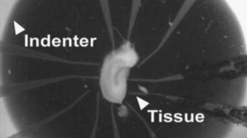
(II) In Vitro Stretch and Compression Injury Models: Traumatic Brain Injury (TBI) & Spinal Cord Injury (SCI)
Mimic stretch and compression injury in vitro using dissociated cell or tissue slice cultures
Perform non-invasive, real-time electrophysiology measurements to directly assess the impact of injury on cellular health and functional activity
Directly compare pre-injury and post-injury electrophysiological data within the same culture for precise functional analysis
Apply multiple, sequential injury events to model repeated trauma, such as mild traumatic brain injury or concussion
Utilize a single system to apply radial, uniaxial, and custom strain profiles, enabling simulation of different injury mechanisms
Ensure highly reproducible mechanical and electrical parameters for consistent, repeatable experiments
Evaluate the neuroprotective efficacy of drug candidates and therapeutic interventions following injury
BMSEED’s stretchable multielectrode arrays (sMEAs) are the only multielectrode arrays compatible with in vitro stretch injury models, uniquely enabling the integration of controlled mechanical injury with simultaneous electrophysiological recording and stimulation.

(III) Cardiac Research
Improve the maturation of cardiomyocytes and promote an adult-like cardiac phenotype by applying controlled biomechanical cues to cell cultures
Generate more predictive in vitro data for cardiac drug efficacy and toxicity, reducing late-stage drug failure and improving clinical trial success rates
Monitor cardiac differentiation and functional maturation using real-time, label-free electrophysiology measurements
Apply precise electrical stimulation for controlled pacing of cardiomyocytes and rhythm modulation
Evaluate cardiotoxicity and proarrhythmic risk of drug candidates early in the development pipeline
BMSEED’s stretchable multielectrode arrays (sMEAs) are the only MEA technology that uniquely combines mechanical stimulation, electrical stimulation, and high-resolution electrophysiological recording in a single integrated system—making them ideally suited for advanced cardiac research and drug screening applications.
(IV) Additional Application Fields for Multielectrode Array (MEA) Technology
Mechanobiology – Study how mechanical forces regulate cellular electrical activity, signaling pathways, and functional behavior
Bottom-Up Neuroscience – Investigate neuronal network formation, connectivity, and emergent electrophysiological behavior from the cellular level
Pain Research – Analyze sensory neuron excitability, neural signaling, and drug response in physiologically relevant in vitro models
Other Emerging Applications – Organ-on-chip models, disease modeling, and custom experimental paradigms
Not sure if multielectrode arrays are right for your research?
Contact BMSEED to discuss whether our cost-effective MEA systems and stretchable multielectrode arrays (sMEAs) can enhance your experimental outcomes and translational relevance.
What Clients Say About our MultiElectrode Arrays
“Tuneable parameters and very fast to use.”
-A. Pybus, Ph.D. Georgia Institute of Technology
“Easiness for acquiring electrical signal in a non-invasive manner.”
-A. Patino, Ph.D. Arizona State University
“The most valuable thing is that we can record from cells multiple times…injure and record on the same device.”
-M.K. Dwyer, M.S. Columbia University

a) organotypic hippocampal slice cultures (OHSCs), long-term potentiation (LTD) & long-term depression (LTD) induced post-injury; b) primary hippocampal neurons (PHNs) & brain tissue tolerance to traumatic brain injury (TBI); c) organotypic spinal cord slice cultures (OSCSCs) & cypin regulated pain sensitivity after spinal cord injury (SCI); d) human induced pluripotent stem cell-derived cardiomyocytes (hiPSC-CMs), hiPSC-CM differentiation; e) neurons, astrocytes, & microglia, pro-inflammatory signaling & immune response to mild traumatic brain injury (mTBI)
Our team of experts...

Founder
Phone: +1 (609) 532-9744
Email: oliver@bmseed.com
MultiElectrode Array Systems
BMSEED’s MEA Electrophysiology Data Acquisition System is available as a stand-alone equipment or integrated with a cell stretcher and imaging capabilities (MEASSuRE platform)

Stand-Alone
120 channel electrophysiology controller
60 or 120 channel recording and stimulation of electrophysiological activity
Stimulation currents from 10 nA - 2.55 mA
Analog and digital stimulation triggers
Compatible with stretchable multielectrode arrays (sMEAs) and glass multielectrode arrays (gMEAs)
Low-cost, high performance data acquisition system for electrophysiology
Contact us for pricing information.

Integrated in MEASSuRE Platform
Mechanics Module: stretching at strain rates up to 90/s and strains up to 50%
Imaging Module: optical or fluorescence imaging during stretching
Electrophysiology Module: 2x60 channels for recording and stimulation
Contact us for pricing information.


Frequently Asked Questions About our MultiElectrode Arrays
1. What is a multielectrode array?
Tools for Electrophysiological Recording and Stimulation
Multielectrode arrays (MEAs)—also known as microelectrode arrays—are advanced platforms used for electrophysiological recording and stimulation of excitable cells and tissues. These microfabricated devices feature a dense grid of microscopic electrodes made from biocompatible, conductive materials such as gold, platinum, or titanium that are deposited onto a a substrate material offering varying degrees of rigidity.
Rigid substrates (e.g., glass, silicon) for high-resolution recordings
Flexible substrates (e.g., polyimide) for conformal tissue interfacing
Stretchable substrates (e.g., PDMS) for studies replicating the mechanically dynamic environment of the human body
At BMSEED, our MEA technologies are engineered to support high-precision research in neuroscience, cardiology, mechanobiology, drug discovery, and tissue engineering. We specialize in both traditional rigid MEAs and innovative stretchable MEA platforms, enabling simultaneous electrophysiology and controlled mechanical stimulation.
Multielectrode arrays offer a versatile tool for studying excitable tissues both in vitro (cultured cells) and in vivo (living organisms). Their primary function involves:
Electrophysiological recording: By virtue of their close proximity to living cells, multielectrode arrays passively capture the minute voltage fluctuations associated with cellular activity (e.g., action potentials, synaptic events) in the extracellular space.
Electrical stimulation: Multielectrode arrays can also be employed to deliver controlled electrical currents to stimulate cells, enabling researchers to probe cellular responses or modulate network activity.
The broad applicability of multielectrode arrays, including both traditional rigid and novel stretchable configurations, allows researchers to investigate diverse biological phenomena, ranging from cellular electrophysiology and network dynamics to drug discovery applications.

2. How does a multielectrode array work?
Multielectrode arrays are critical components in the process of acquiring electrical signals from cells. We describe briefly the process of how multielectrode arrays are used in recording and stimulation of electrophysiological activity.
Cellular Interface: The multielectrode array surface is typically composed of biocompatible materials and presents a microfabricated grid of microelectrodes (e.g., gold, platinum) for intimate contact with cultured cells. This biocompatibility enables acute and long-term (months) electrophysiological studies.
Extracellular Recording: As excitable cells (neurons, cardiomyocytes) adhere and form networks on the MEA, their electrical activity (action potentials, synaptic events) generates minute voltage fluctuations (typically 10s to 100s of μV) in the extracellular space.
Signal Transduction: These voltage changes are passively captured by the electrodes due to their close proximity to the cells. The microelectrodes on the multielectrode array basically transduce ionic currents in the medium that are caused by cellular electrical activity into electronic currents.
Signal Processing: The acquired signals are weak and require amplification to enhance their amplitude for further analysis. The amplifiers on the headstage amplify the differential signal of recording and reference electrodes. The amplifiers on BMSEED’s MEA electrophysiology system are placed in proximity to the electrodes to minimize noise.
Noise Elimination: Filtering techniques are employed to eliminate unwanted electrical noise originating from the environment or the multielectrode array itself. BMSEEED’s multielectrode array electrophysiology system has a notch filter to eliminate 50Hz and 60Hz line noise, as well as low-pass filters and high-pass filters with adjustable frequencies.
Data Acquisition: The amplified and filtered signals are then digitized and transferred to a computer system for further processing and analysis.
Electrophysiological Analysis: Specialized software allows researchers to analyze the recorded activity, including spike sorting (identifying individual neuron activity), network analysis (evaluating communication patterns), and field potential characterization (understanding overall network behavior). Ask BMSEED for the NeuroExplorer software.
Multielectrode arrays offer a minimally invasive approach for studying large ensembles of excitable cells, providing valuable insights into cellular electrophysiology, network dynamics, and drug discovery applications.
-
Multielectrode arrays (MEAs) offer several key benefits for electrophysiological measurements:
Network Analysis: MEAs record from multiple electrodes simultaneously, enabling researchers to gather data from a large number of cells or across a larger tissue area compared to single-electrode techniques. This capability allows researchers to analyze the communication patterns and synchronization within neural networks or other excitable cell cultures.
Label-Free: MEAs enable the recording of electrophysiological signals without the use of any labeling dyes (e.g., voltage sensitive dyes). These dyes, e.g., voltage-sensitive dyes, may affect how the cells function and thus the results of the experiment. Using MEAs avoids these issues.
In-Situ, Real Time: The presence of the multiple electrodes on an MEA does not affect the physical environment of the cells for in vitro analysis. The electrophysiological data are displayed in real time.
No Tissue Damage: In vitro studies using MEAs avoid the need for insertion of electrodes into the tissue, thus reducing potential damage to the tissue.
Extracellular Recording: MEAs capture the electrical activity of cells from the surrounding extracellular space, offering a non-invasive alternative to intracellular patch-clamp recording.
Drug Discovery: The ability to monitor cellular activity in response to drugs or compounds makes MEAs valuable tools for drug screening and development.
MEAs provide a powerful and versatile platform for studying cellular electrophysiology and network dynamics, offering significant advantages over traditional single-electrode techniques.
-
Multielectrode arrays record the electrical activity from the extracellular space of a population of cells (neurons, muscle cells) whereas patch clamp electrophysiology records the action potential electrical activity from the intracellular space of a singlecell.
Advantages
Recording data points from a network/population of cells simultaneously
Analysis of cellular network activity and synchronization
Coverage of a large area (several millimeter)
Non-invasive
Relatively easy to use
Relatively inexpensive
Disadvantages
Lower signal to noise ratio than patch-clamp.
-
BMSEED is the only company that offers stretchable multielectrode arrays (sMEAs) for in vitro research and drug development applications. Below we outline the advantages of using stretchable multielectrode arrays for in vitro studies.
Mimicking in vivo conditions: In vitro studies ultimately aim to predict in vivo behavior. There are many reasons for using in vitro studies instead of carrying out in vivo studies directly. Compared to in vivo studies, in vitro studies provide: (a) reduced cost, (b) increased speed, (c) tight control of chemical and physical environment, (d) higher throughput, (e) reduced animal-use, and (f) evaluation of biological phenomena without the potential confounding variables present in whole organisms. The drawback of in vitro studies is that the cellular environment is often very different from the environment in vivo that the cells experience. This difference causes cells to often behave differently in vivo than in vitro. BMSEED’s stretchable multielectrode arrays aim to reduce the difference between in vivo and in vitro cellular environments by replicating biophysical aspects of the living tissue environment in a controlled environment in vitro.
Improved cell viability: Rigid multielectrode arrays can exert pressure on cultured cells, potentially affecting their health and viability. The conformability of stretchable multielectrode arrays can minimize this stress, leading to healthier and more representative cell cultures for in vitro studies.
Studying dynamic cell cultures: Certain in vitro models involve culturing cells on flexible substrates that mimic specific tissues or organs. Stretchable multielectrode arrays can better integrate with these dynamic environments, maintaining good contact with the cells as they move or deform.