
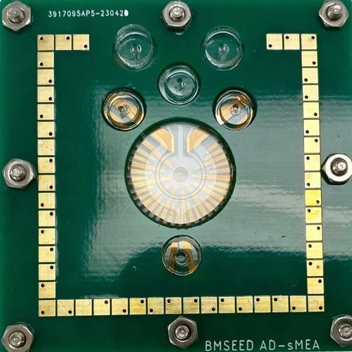
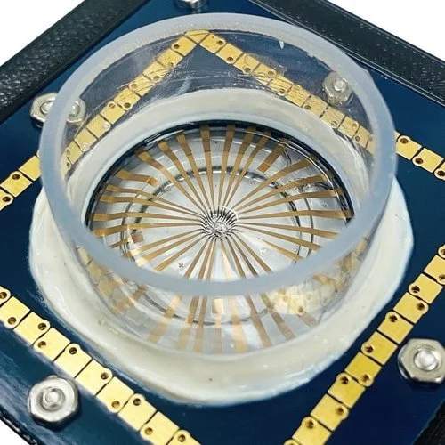
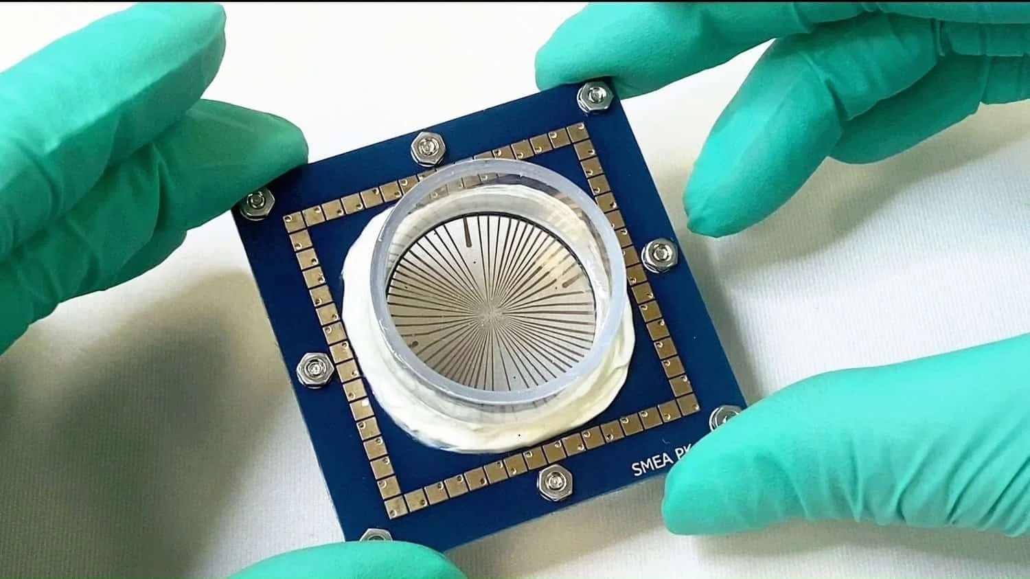
Microelectrode Array
✓ Record electrophysiological activity across various locations within the culture
✓ Reproduce biophysical cues in vitro
✓ Best value MEA electrophysiology data capture system
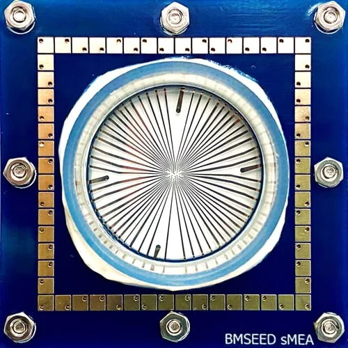
A key drawback of in vitro experiments is their inability to fully replicate the natural environment of cells within a living organism. The accuracy of predictions from in vitro data diminishes significantly when primary cells are isolated and cultured on rigid MicroElectrode Arrays (MEAs), which differ substantially from how these cells behave in vivo. To overcome this issue, BMSEED’s stretchable MicroElectrode Arrays (sMEAs) incorporate flexible electrodes integrated into an elastomeric material. These electrodes interact with cell or tissue cultures, simulating the mechanical and electrical conditions found in vivo while maintaining a controlled in vitro environment. sMEAs help bridge the divide between in vivo and in vitro research by allowing for mechanical extension, optical visualization, and electrical stimulation and recording, thus improving the relevance and applicability of experimental findings.
Why Our MicroElectrode Arrays?
✓ Lower cost & simplified set up compared to alternative electrophysiological techniques
✓ In-situ, label-free, and real-time measurements
✓ 2D and 3D cell cultures, slices, and organoids
✓ 30, 60, & 120 channels
✓ Electrical stimulation
✓ Acute & chronic experiments
✓ Reproduce biophysical in vivo environment
✓ Largest variety of microelectrode arrays (2D, 3D, soft, traditional)
✓ In vitro results are more predictive of in vivo behavior
✓ Evaluate drug candidate efficacy and toxicity

Our dedicated team of specialists...
Founder
Email: oliver@bmseed.com
Phone: +1 (609) 532-9744
Fields of Application of MicroElectrode Arrays

(I) Improve the Prediction of In Vivo Behavior Using In Vitro Data
Soft substrates, unlike glass or plastic, mimic the in vivo microenvironment
Reproduce the bio-electrical and bio-mechanical and in vivo cellular surroundings
Compatible with human-derived iPSCs
Consistent and reproducible data with precise regulation of experimental conditions.
Our stretchable microelectrode arrays are the EXCLUSIVE MEAs that combine the implementation of mechanical stretch using electrophysiological assessments

(II) Neurotrauma: Traumatic Brain Injury (TBI) and Spinal Cord Injury (SCI)
Reproduce injury biomechanics in vitro
Evaluate the effects of injury on the condition and functionality of cells using BMSEED’s non-invasive electrophysiology module
Normalize post-injury electrophysiological data to pre-injury baseline measurements
Examine the effects of multiple injuries, e.g., repeated concussions
Examine various types of injuries by applying diverse strain profiles (radial, uniaxial, and others) using a single setup
Obtain reproducible data with highly reproducible experimental parameters
Evaluate neuroprotective treatments
Our stretchable microelectrode arrays are the EXCLUSIVE compatible MEAs with mechanical injury models.
(III) Cardiac
Produce cardiac cells with a mature adult-like phenotype by applying biomechanical stimuli to the culture
Reduce the failure rate of clinical trials by more accurately assessing the effectiveness and safety profile of drugs in vivo using in vitro data
Monitor differentiation and development of cardiomyocytes (and iPSC-derived neurons) in vitro with label-free, real-time electrophysiology measurements
Apply regulated electrical stimulation to pace cardiomyocyte activity
Evaluate drug candidates for cardiotoxicity and proarrhythmic effects
Our stretchable microelectrode arrays are the EXCLUSIVE MEAs that enable the concurrent and independent application of cell cultures stimulated mechanically and electrically in conjunction with electrophysiological measurements.
(IV) Additional Areas of Application for MicroElectrode Arrays
Pain research
Mechanobiology
Bottom-up neuroscience
other
Contact us to discuss how our stretchable microelectrode arrays might benefit your research.
What Customers Say About our MicroElectrode Arrays
“Tuneable parameters and very quick to use.”
-Dr. Pybus, Georgia Institute of Technology
“Simplicity in obtaining electrical signals in a non-invasive way…”
-Dr. Patino, Arizona State University
“The greatest advantage is the ability to repeatedly record from the same cells, allowing for injury and subsequent recordings on a single device.”
-M. K. Dwyer, M.S. Columbia University

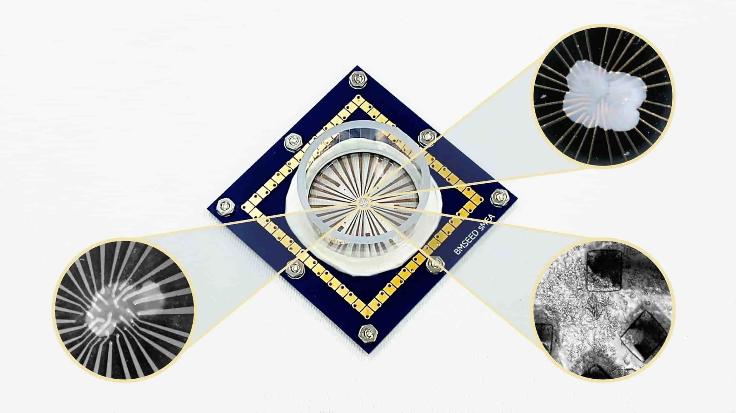
a) Organotypic hippocampal slice cultures (OHSCs) with post-injury induction of long-term potentiation (LTP) and long-term depression (LTD); b) Primary hippocampal neurons (PHNs) and brain tissue resilience to traumatic brain injury (TBI); c) Organotypic spinal cord slice cultures (OSCSCs) and cypin-mediated regulation of pain sensitivity following spinal cord injury (SCI); d) Human-induced pluripotent stem cell-derived cardiomyocytes (hiPSC-CMs) and the differentiation process of hiPSC-CMs; e) Neurons, astrocytes, and microglia, focusing on pro-inflammatory signaling and immune response following mild traumatic brain injury (mTBI).
Our dedicated team of specialists...

Founder
Phone: +1 (609) 532-9744
Email: oliver@bmseed.com
MicroElectrode Array Systems
Use stretchable microelectrode arrays (sMEAas) as well as traditional 2D and 3D microelectrode arrays with BMSEED’s MEA System for Electrophysiology Data Capture platform either as an individual instrument or incorporated with a cell stretcher and imaging equipment (MEASSuRE platform).
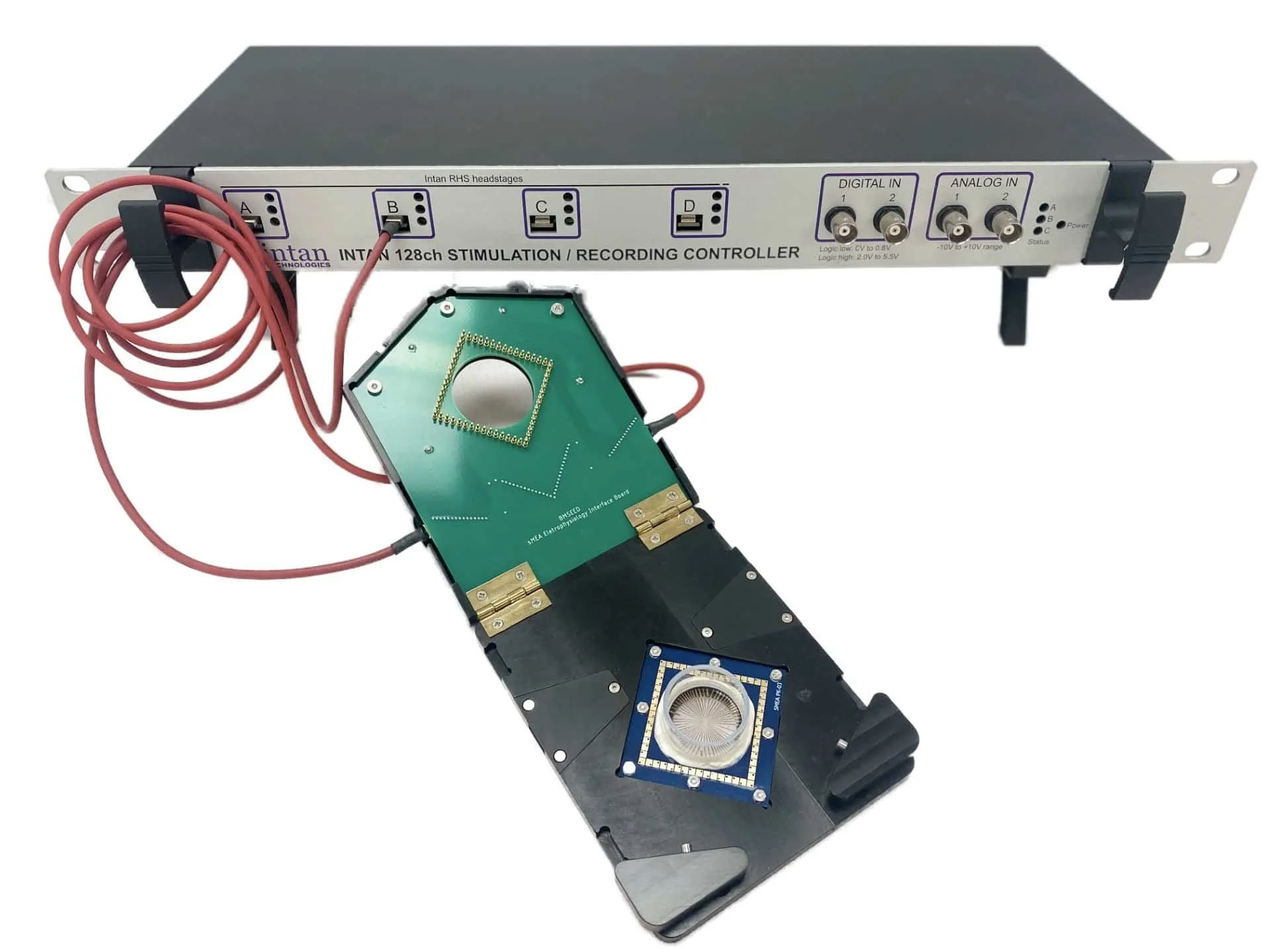
Stand-Alone
120 channel MEA electrophysiology controller
Monitoring and stimulating electrophysiological signals activity using MEAs with 30, 60, or 120 microelectrodes
Apply stimulation currents from 10 nA up to 2.55 mA
Apply analog or digital signal triggers, e.g., audio, visual, function generator
Use the same equipment for stretchable microelectrode arrays (sMEAs) and glass microelectrode arrays (gMEAs)
State-of-the-art advanced data capture platform for electrophysiological studies in the industry
Reach out to inquire about pricing details
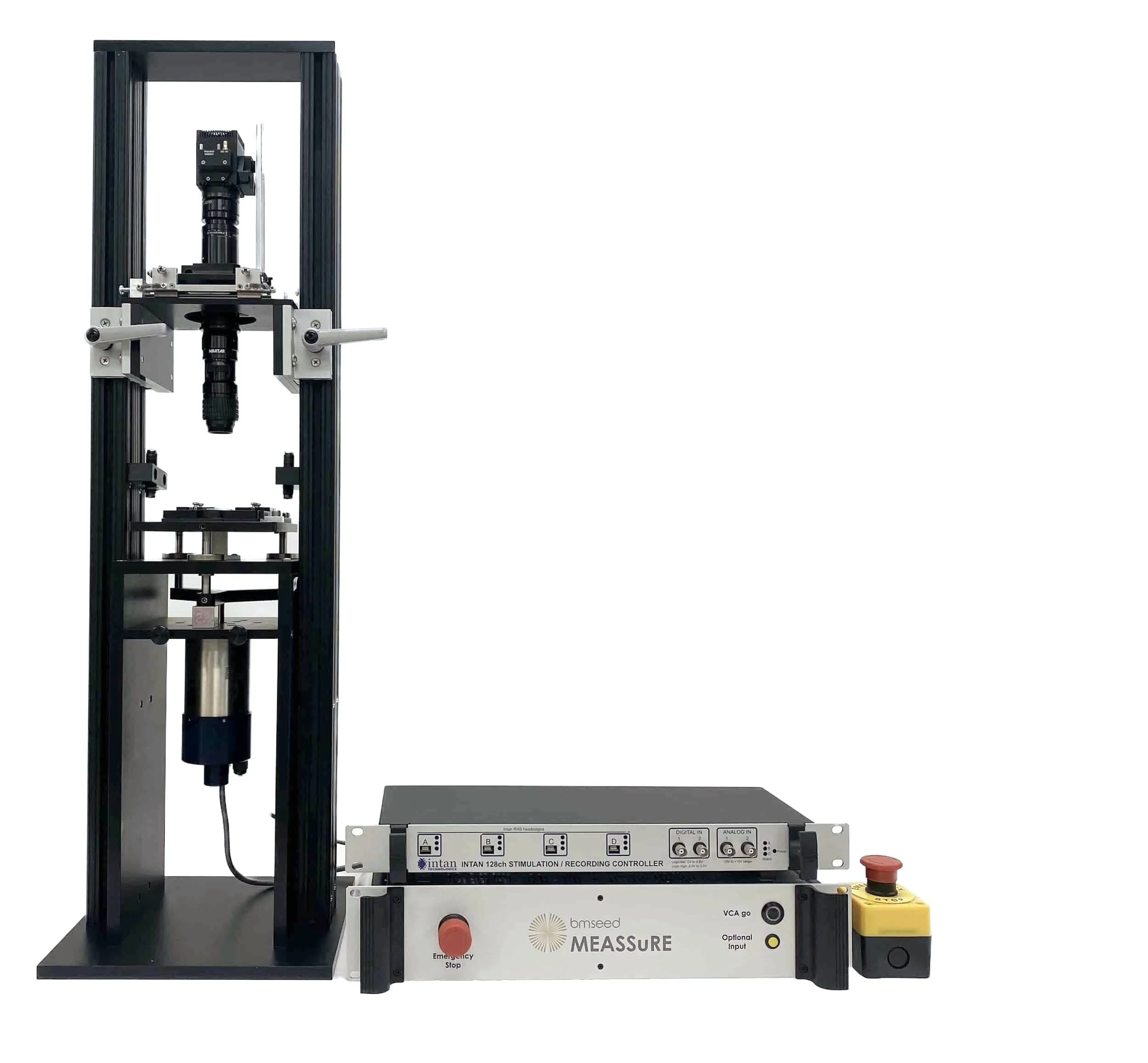
Integrated with MEASSuRE Platform
Mechanics Module: mechanical stretching at high strain rates of up to 90/s and strains of up to 50%
Imaging Module: visualize cell during stretching using optical or fluorescence imaging
Electrophysiology Module: 120 measurement and excitation channels
Reach out to inquire about pricing details
60/120 channels
Stiff & inflexible
Recording & stimulation
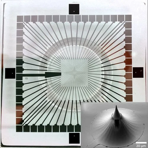
3D (small spikes)
Organoids, slices
Common Questions Regarding Our MicroElectrode Arrays
1. What is a micro-electrode array?
Devices for Recording and Stimulating Electrophysiological Activity
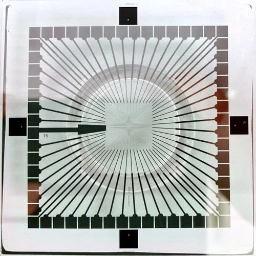
Microelectrode arrays (MEAs), aka multielectrode arrays, are microfabricated devices for electrophysiological measurements, both in vivo (living organisms) and in vitro (cultured cells). MEAs consist of an array of small electrodes that are made of electrically conductive and biocompatible materials, including gold, platinum, and titanium. The substrate materials that the electrodes are deposited onto offer range from rigid materials (glass) to flexible (polyimide) and stretchable (PDMS).
The main purpose of MEAs includes:
Electrophysiological monitoring: MEAs, paired with a data capture system, detect minute voltage changes linked to cellular activity (such as action potentials and synaptic activities) within the extracellular environment.
Electrical stimulation: The electrodes on MEAs can apply precise electrical impulses to activate cells, allowing the study of cellular responses and network dynamics.
MEAs provide critical information for a broad range of biological applications, spanning cellular electrical activity and neural networks to pharmaceutical development.
BMSEED’s traditional MEAs and stretchable MEAs, both in 2D and 3D configurations, provide a versatile tool set for researchers to overcome technology limitations.
2. How does a micro-electrode array work?
Microelectrode arrays are a key component in acquiring electrical signals from cells. We briefly describe below how MEAs facilitate both the detection and induction of electrophysiological signals.
Cellular Interface: The MEA surface is composed of a microfabricated grid of microelectrodes made from biocompatible materials (e.g., gold, platinum) that are in intimate contact with cultured cells. With biocompatibility, both acute and sustained (months) electrophysiological investigations are possible.
Extracellular Recording: Excitable cells such as neurons and cardiomyocytes produce electrical action potentials that generate small variations in voltage (usually ranging from tens to hundreds of μV) within the extracellular environment.
Signal Transduction:The MEA’s microelectrodes convert ionic currents from the cellular electrical activity in the medium into electronic signals.
Signal Processing: These weak signals are amplified to enhance their amplitude by amplifiers on the headstage, which are positioned near the electrodes to reduce the pickup of electrical noise.
Noise Elimination: To mitigate unwanted electrical noise, notch filters can be used to remove 50Hz and 60Hz line signals, while adjustable low-pass and high-pass filters offer additional filtering options.
Data Capture: The digitized amplified and filtered signals are transferred to a computer system for signal processing.
Electrophysiological Analysis: The analysis of the recorded electrophysiological activity (spike sorting, network analysis, field potential characterization) is either done by custom codes specific to a lab or by specialized software. Contact BMSEED to request the NeuroExplorer software.
MEAs provide a non-invasive, in-situ methodology for studying groups of excitable cells that provide valuable insights into cellular electrophysiology and network dynamics.
-
Microelectrode arrays (MEAs) offer numerous advantages for electrophysiological measurements compared to other techniques:
Network analysis: MEAs record electrical signals simultaneously with multiple electrodes from a large number of cells and across a larger area compared to single-electrode techniques, which enables the analysis of communication patterns and synchronization within neural networks or other excitable cell cultures.
Label-free: MEAs do not require labeling with dyes for recording electrophysiological signals. These dyes, e.g., voltage-sensitive dyes, may affect how the cells function and thus the results of the experiment, which is highly undesirable. Using MEAs avoids these issues.
In-Situ, real time: The electrodes on an MEA do not affect the physical environment of the cells for in situ measurements, and the electrophysiological data are recorded and displayed in real time.
No tissue damage: In vitro MEAs are not inserted into the tissue, thus avoiding potential tissue damage.
Extracellular recording: MEAs capture the electrical activity of cells at multiple locations simultaneously from the extracellular space, providing a powerful and non-invasive alternative to patch-clamp methods.
Drug discovery: MEAs are valuable tools for drug development because of their ability to monitor cellular activity in response to drugs, toxins, and other compounds.
MEAs provide a powerful and versatile platform for studying cellular electrophysiology and network dynamics, offering significant advantages over other techniques.
-
MEAs capture the electrical signals from the extracellular space from a collection of cells (neurons, muscle cells) while patch clamp captures action potentials from the intracellular space of a single cell.
Advantages
Capture signals from a network/population of cells simultaneously and concurrently
Enable analysis of network activity and synchronization
Coverage of a large area (several millimeter)
Non-invasive method
Relatively easy to use compared to patch-clamp
Relatively inexpensive compared to patch-clamp
Disadvantages
Lower signal to noise ratio than patch-clam
-
Stretchable microelectrode arrays (sMEAs) for in vitro research and drug development applications are only offered by BMSEED. Below we outline the advantages of using sMEAs for in vitro studies.
Mimicking in vivo conditions: In vitro studies aim to predict in vivo behavior. There are numerous reasons for using in vitro studies instead prior to or instead of in vivo studies. In vitro studies provide the following benefits compared to in vitro methods: (a) lower cost, (b) higher speed, (c) control of chemical and physical environment, (d) higher throughput, (e) fewer animals are required, and (f) absence of confounding variable that are present in whole organisms. The disadvantage of in vitro studies is that the cellular environment is different from the cellular environment in vivo, which can cause cells to behave differently. Our stretchable MEAs aim to reduce the difference between in vivo and in vitro cellular environments by replicating biophysical aspects of the living tissue environment in a controlled environment in vitro.
Improved cell viability: Cells sense the rigidity of the substrates they grow on, thus behaving differently on rigid compared to soft substrates. The softness and compliance of stretchable MEAs can minimize these stresses, leading to healthier and more representative cell cultures for in vitro studies.
Studying dynamic cell cultures: Some in vitro models that aim to mimic specific tissues or organs use flexible substrates for culturing cells. Stretchable MEAs have an additional degree of flexibility, thus maintaining good contact with the cells as they move or deform.