
3D Multielectrode Arrays (3D-MEAs)
MultiElectrode Arrays are Consumables for MEASSuRE: View Catalog
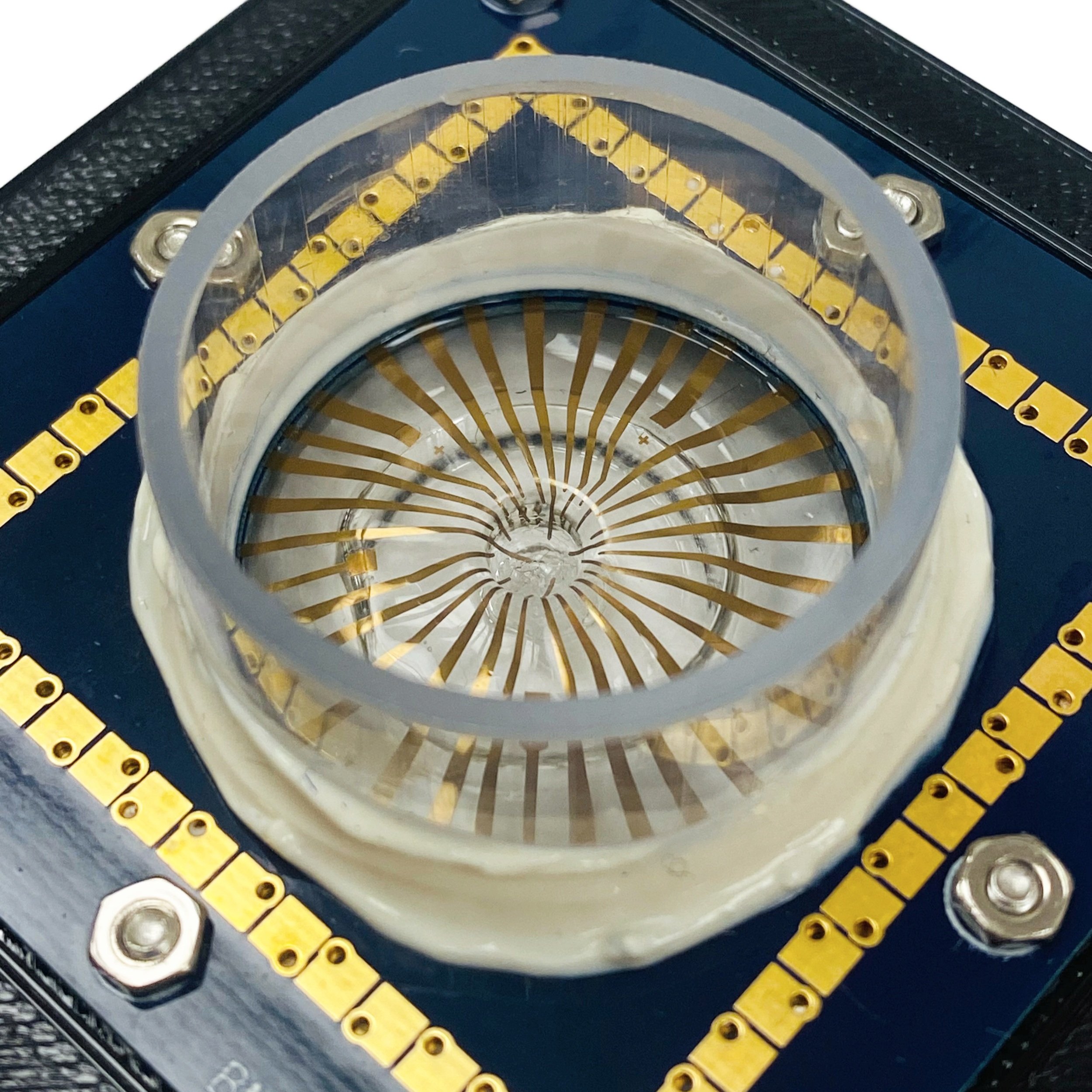
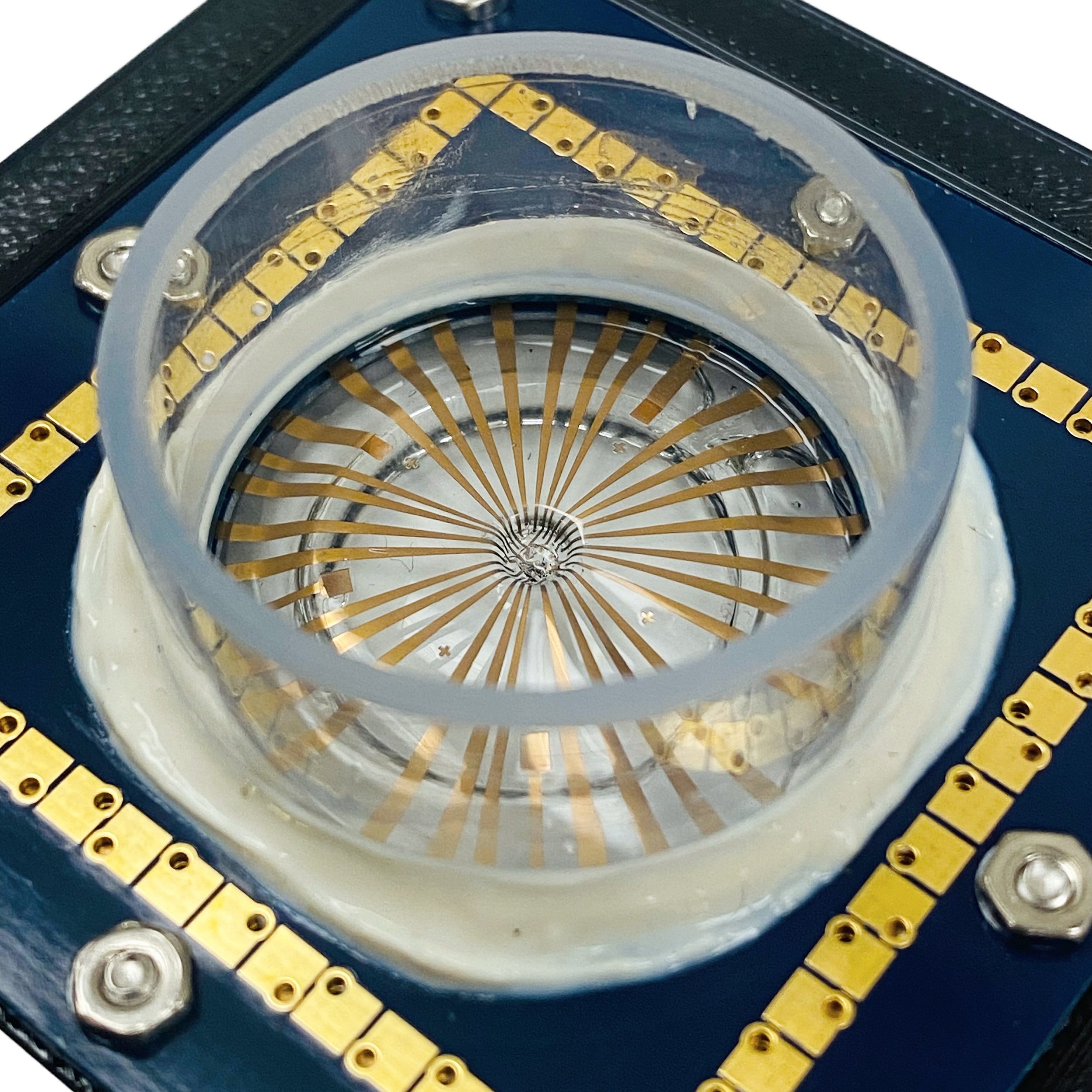
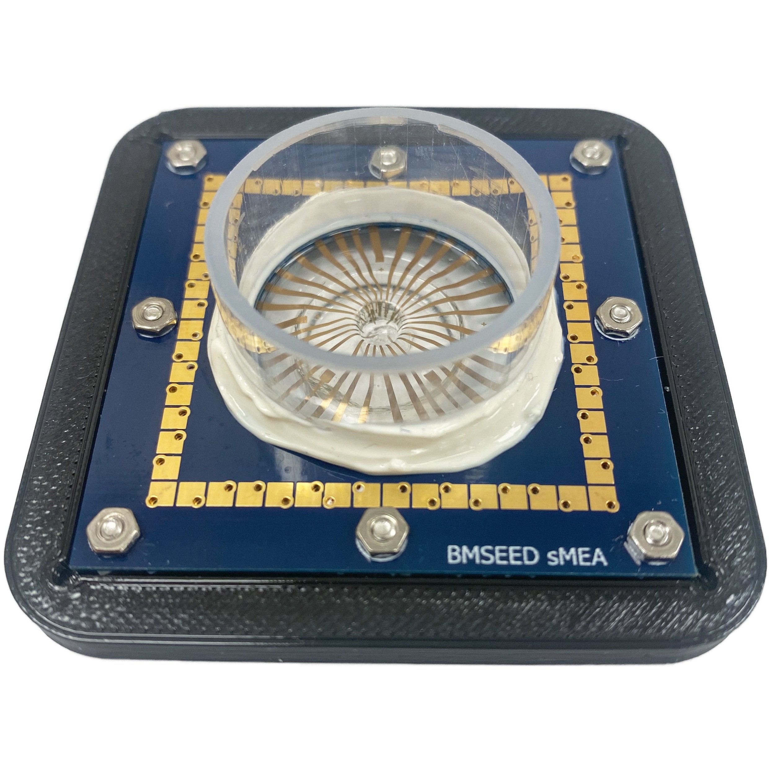
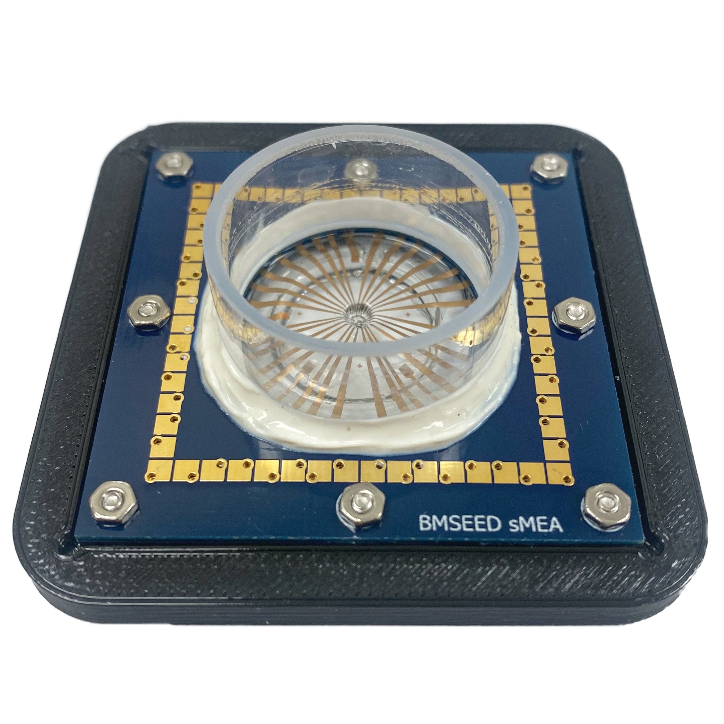
BMSEED’s 3D multielectrode arrays (3D-MEAs) are designed for organoid and cell culture research, allowing accurate electrophysiological recording and stimulation on a stretchable silicone substrate. The system includes a 3D pocket to hold organoids, maintaining shape for enhanced physiological relevance and synchronized network measurements. Customizable in size (1-5 mm diameter, 0.8-2 mm depth), the pocket supports various organoid types. For cell cultures, the dual-chamber microfluidics enable co-culturing and media delivery. With customizable electrode layouts, the 3D-MEAs are ideal for advanced applications in neurobiology, mechanotransduction, and tissue engineering.
In Vitro Research
32pMEA-100-300-4iR-Au-1W and 60pMEA-100-420-4iR-Au-1W
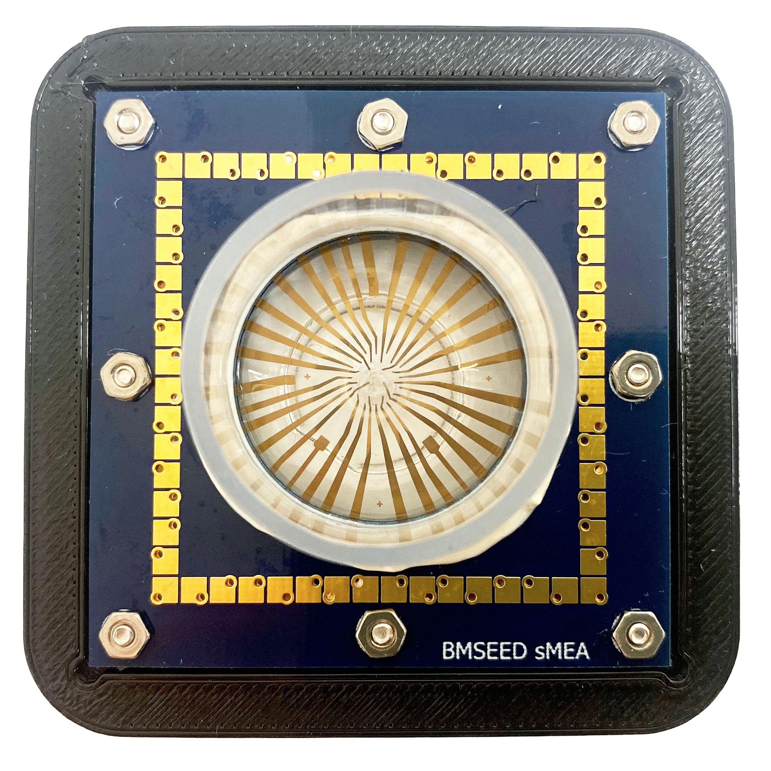
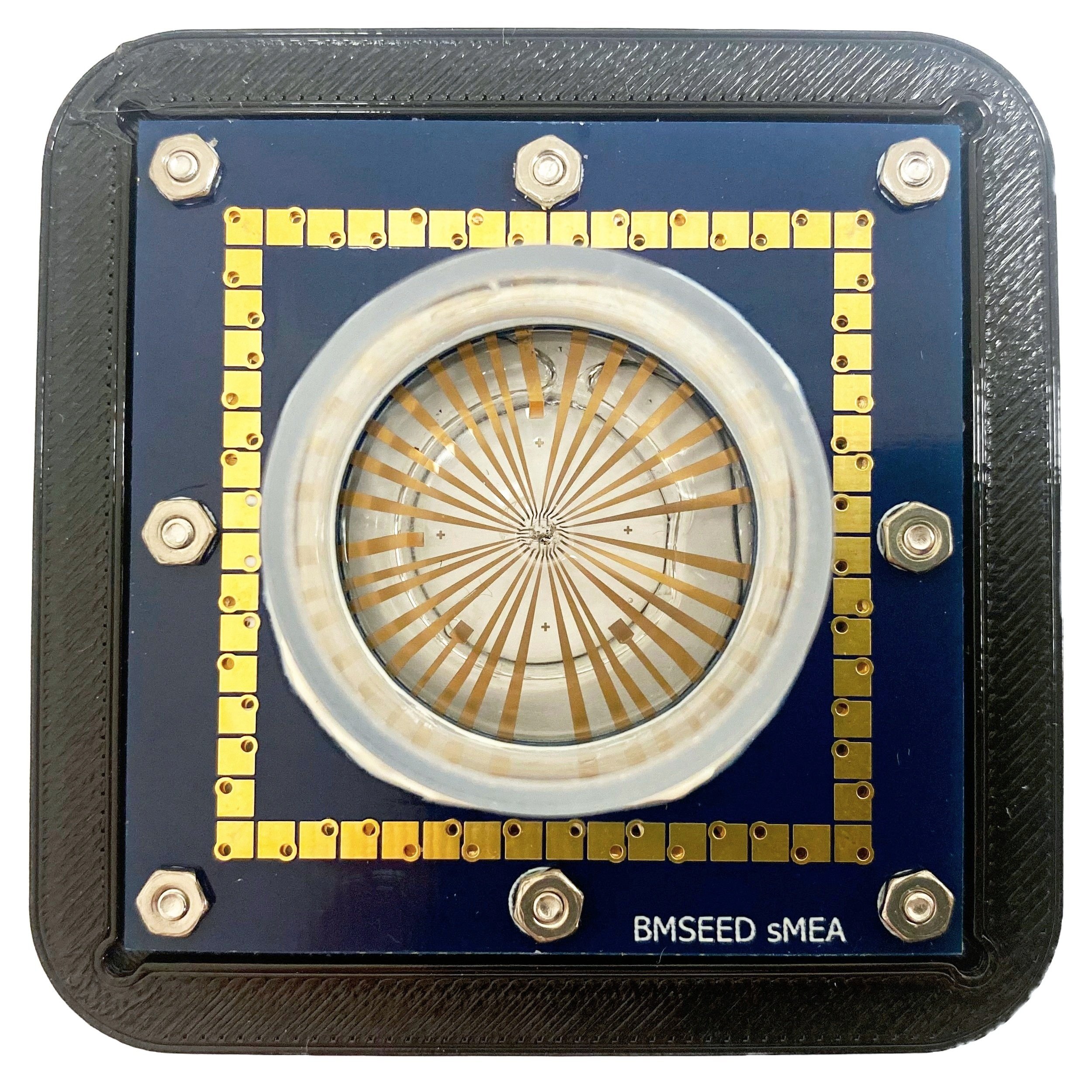
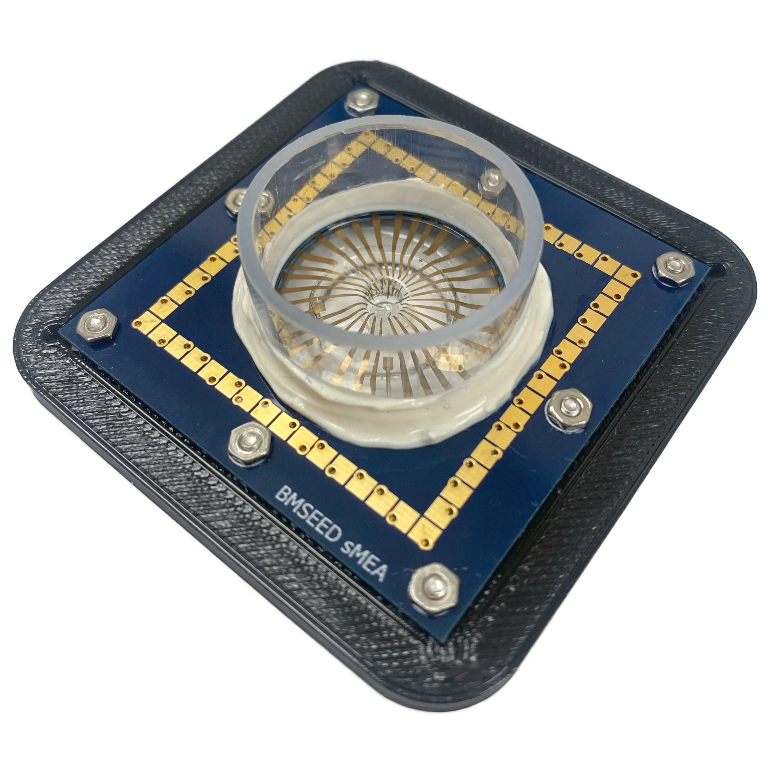
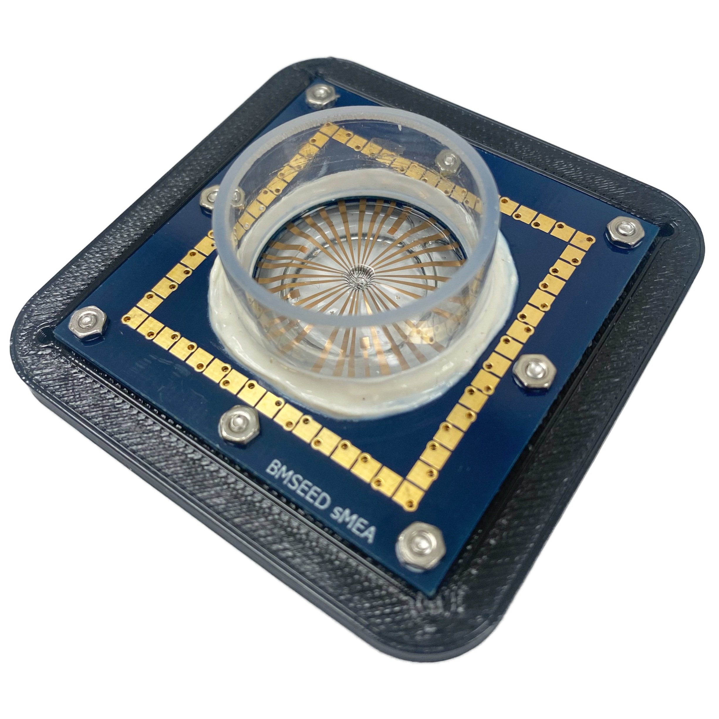
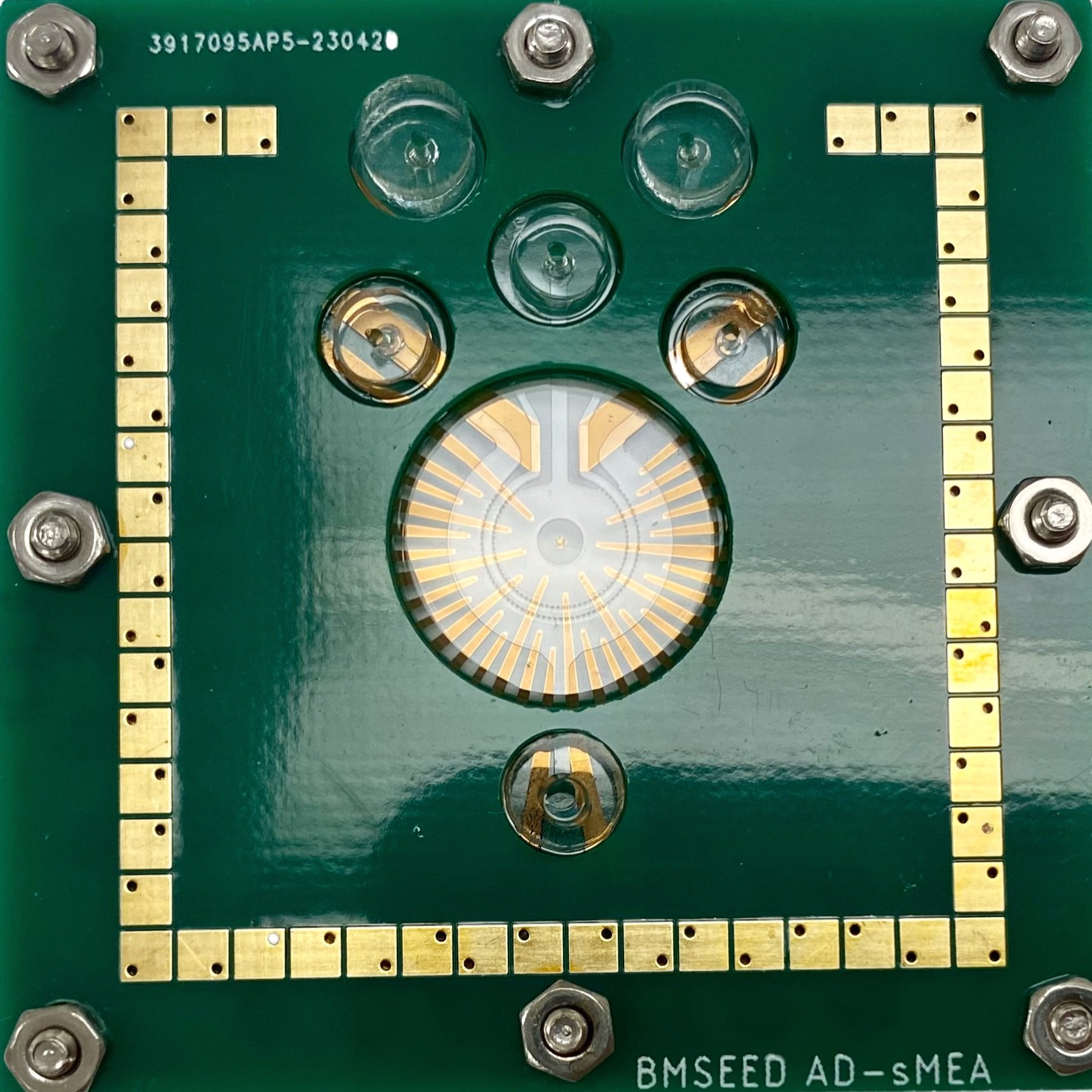
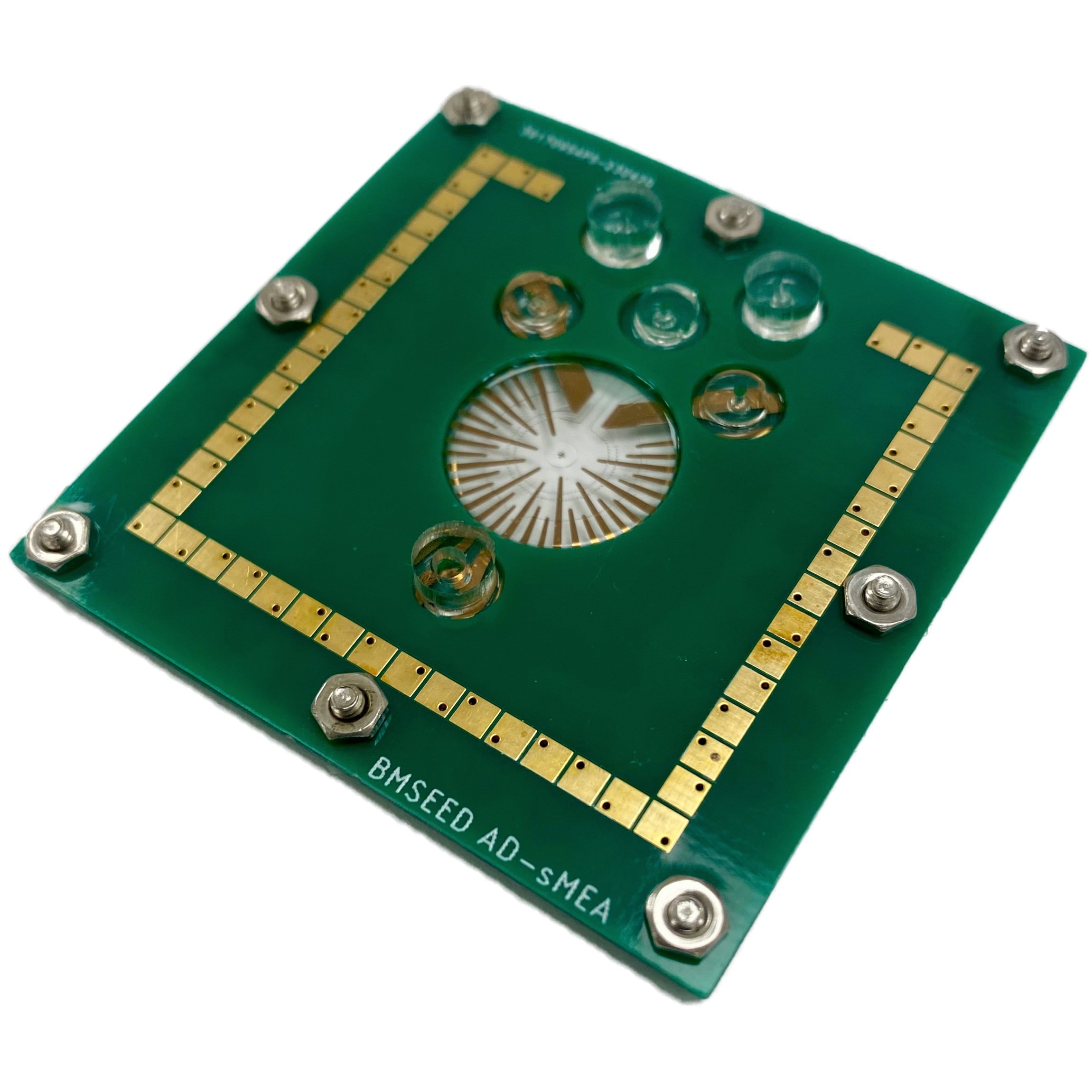
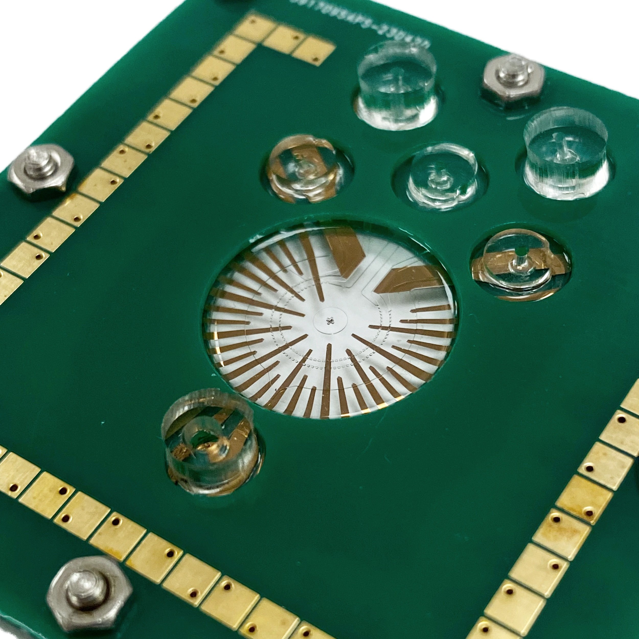
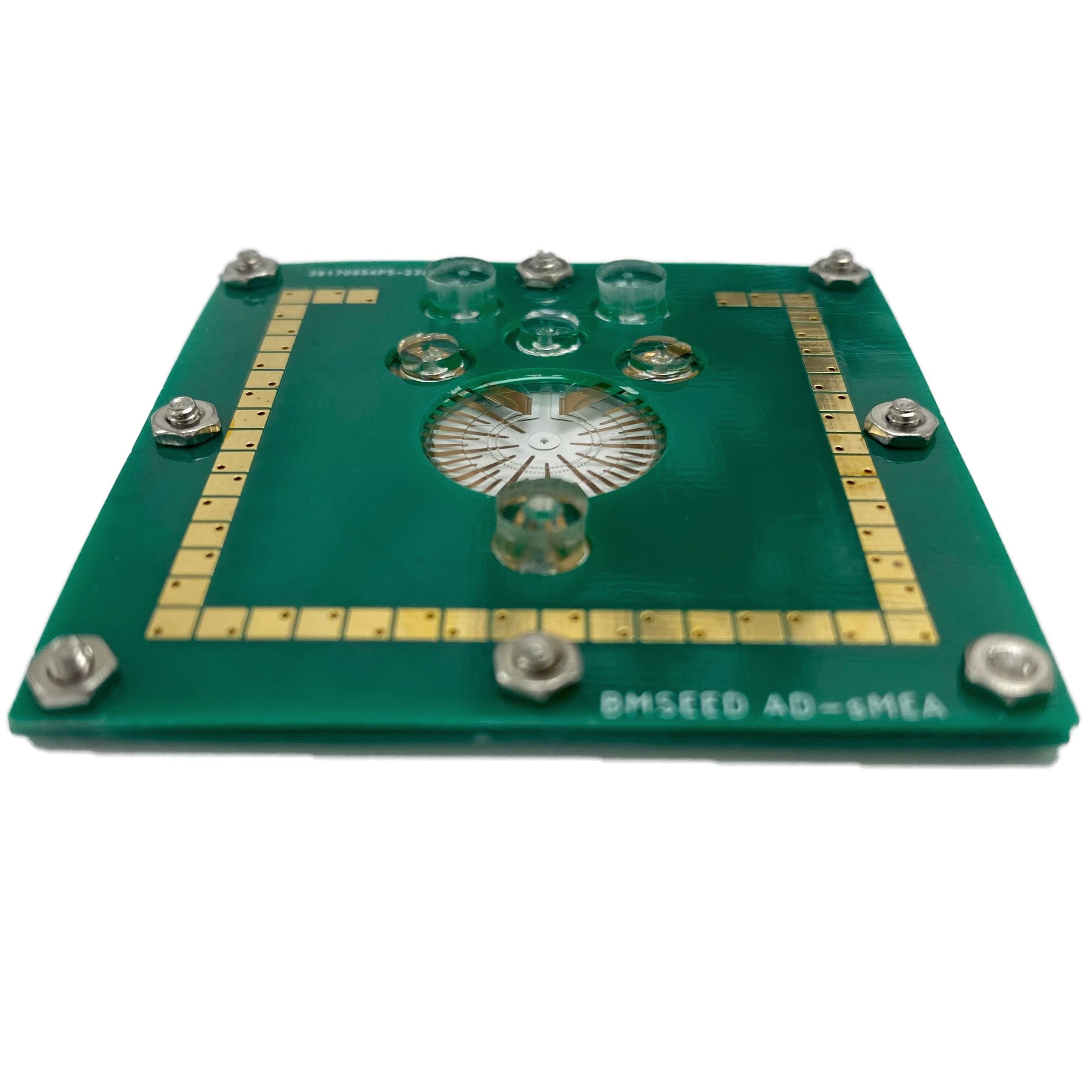
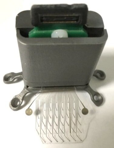
Organoid MEA: Stretchable multielectrode array with fixed 3D Pocket
Features and Benefits
organoid does not remodel in pocket
retain organoid shape
increased physiological relevance
recording from a large surface area of the organoid
more accurate network synchronization measurements
recording & stimulation of electrophysiological activity
silicone substrate & pocket
customizable pockets: 1-5 mm diameter, 0.5-1.5 mm depth
customizable electrode layouts
product number (PN): 11-53100 (32-channel), 11-53200 (60-channel)
discounted units available for early adopters
Stretchable microelectrode array with 3D pocket for organoid research
Image (lower left): cerebral organoid in BMSEED’s sMEA with 3D pocket, J. Guo, Emory University
34mMEA-200-960-4iR-Au-1W
Microfluidic stretchable multielectrode array with dual-chamber for3D Cell Cultures
Features and Benefits
dual-chamber for co-culturing
recording & stimulation of extracellular electrophysiological activity
elastically stretchable (silicone) substrate & microstructures
compatible with perfusion systems to deliver cell media
customizable patterns & number of electrodes
product number (PN): 11-54100
Image: neurons seeded in BMSEED’s 3D-SW with microchannels. A. Palmieri, B.S., Georgia Institute of Technology
Cell Cultures: stretchable multielectrode array with microchannels for neural axon growth
promote structured neuronal networks with axonal guidance to study how neuronal structure correlates to function
recording & stimulation of extracellular electrophysiological activity
elastically stretchable (silicone) substrate & microstructures
customizable patterns & number of electrodes
Image (right): cortical rat neurons seeded in BMSEED’s sMEA with microchannels, ETH Zurich.
In Vivo Brain Research
Soft and compliant electrodes for electrocorticography (ECoG) in brain research applications
ECoG array with printed circuit board and ZIF clip connector
ECoG MultiElectrode Array
65 microelectrodes + 4 reference electrodes
large area & high resolution
recording & stimulation of extracellular electrophysiological activity
elastically stretchable (silicone substrate)
soft
product number (PN): 11-56110
Pedestal to securely mount the ECoG on the skull without hurting the animal
In Vivo Multielectrode Array for Peripheral Nerve Research
Soft and compliant electrodes for interfaces with the peripheral nervous system (PNS)
PNI with printed circuit board
Peripheral Nerve Interface (PNI)
6 microelectrodes
recording & stimulation of extracellular electrophysiological activity
elastically stretchable (silicone substrate)
soft
product number (PN): 11-56210
Printed microclip to anchor the PNI to any nerve size without sutures or surgical adhesives

SELECTION GUIDE FOR CONSUMABLE
Identify whether to use stretchable multielectrode arrays, stretchwells (no electrodes), or glass multielectrode arrays.